
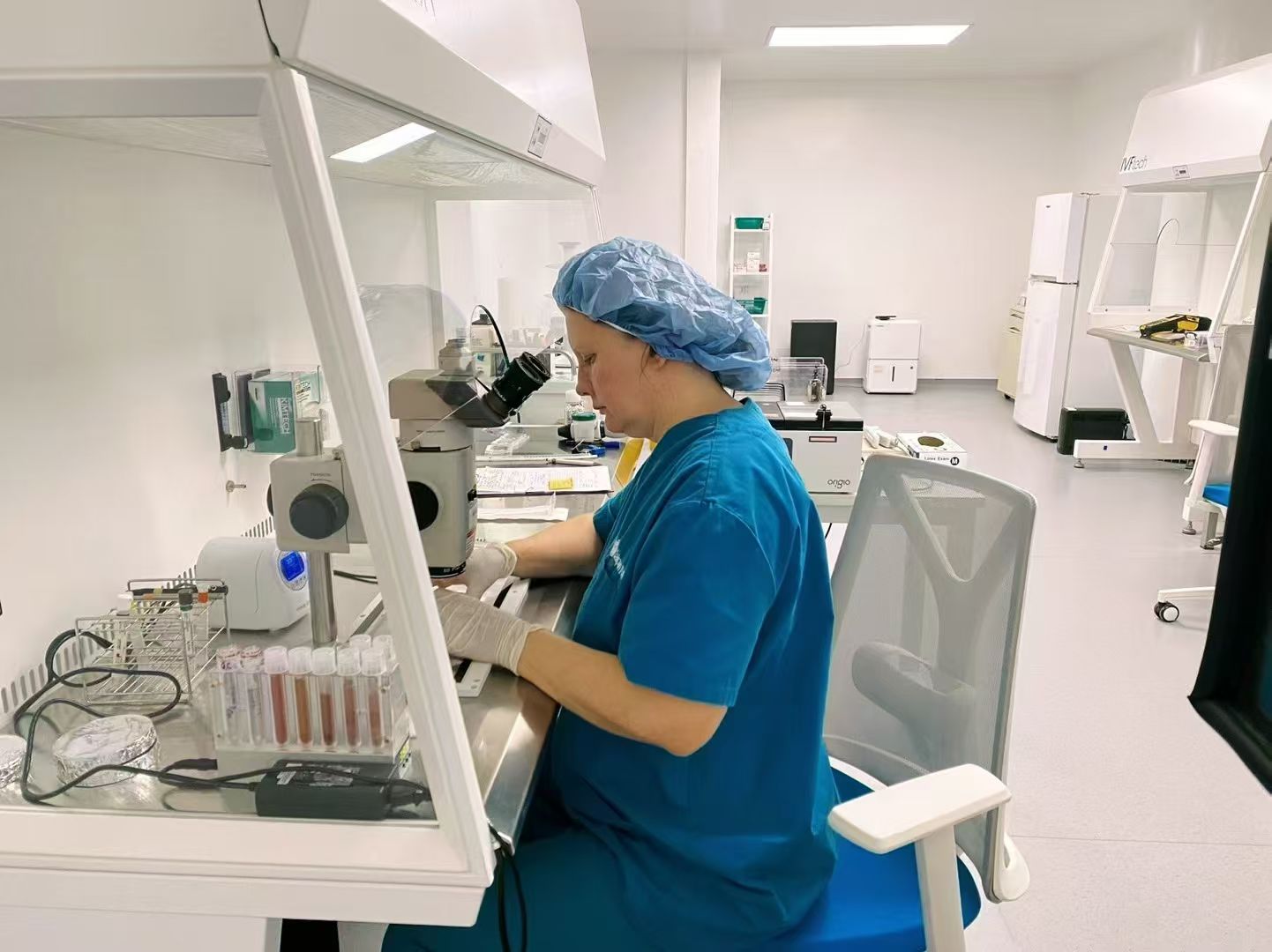
Once egg retrieval is over, the most closely watched part of an IVF cycle happens somewhere the patient never goes: the embryology laboratory. Over the next five to six days, eggs are inseminated, checked for fertilization, cultured in incubators, and assessed as they divide from a single fertilized cell into a blastocyst of a hundred cells or more. Patients experience this week mostly as a series of phone calls, beginning with the fertilization report, and as a stream of unfamiliar vocabulary: pronuclei, cleavage stage, morula, blastocyst, and grades that read like seat assignments, such as 4AA. This article walks through what happens to eggs and sperm immediately after retrieval, when and how the fertilization report is shared, how an embryo develops day by day, the difference between a day 3 embryo and a day 5 blastocyst, how cleavage-stage and blastocyst grading systems work, why some embryos stop developing, and which questions are worth bringing to the clinic. It draws on patient information from the American Society for Reproductive Medicine's ReproductiveFacts.org, Guy's and St Thomas' NHS Foundation Trust, Cleveland Clinic, the UK fertility regulator HFEA, an open-access review of the SART embryo grading system, Illume Fertility, and CCRM Fertility. It is general education only, and every individual decision belongs with the treating clinic and a licensed physician.
What happens to eggs and sperm right after egg retrieval?
The laboratory takes over the moment the follicular fluid leaves the procedure room. The ASRM ReproductiveFacts.org booklet explains that mature eggs are placed in an IVF culture medium and transferred to an incubator to await fertilization by the sperm. On the same day, the sperm sample is prepared: Guy's and St Thomas' NHS Foundation Trust describes putting the normal and moving sperm into an electronically tagged test tube, part of the witnessing systems laboratories use to keep every sample matched to the right patient. Insemination then happens in one of two ways. In conventional IVF, ASRM describes motile sperm being placed together with the oocytes and incubated overnight, and Guy's and St Thomas' similarly describes sperm and eggs being put together in a carefully labelled and electronically tagged dish and left in an incubator overnight. In ICSI, ASRM explains that a single sperm is directly injected into each mature egg, and Guy's and St Thomas' notes that in an ICSI cycle each egg is injected individually with a single sperm, an approach clinics may recommend when sperm count or motility is low. Which method is used for any given cycle is a clinical and laboratory decision made case by case, so anyone unsure why conventional IVF or ICSI was chosen for them should put that question directly to the treating clinic and a licensed physician.
What is a fertilization report and when do patients usually hear about it?
The fertilization report is the first news from the laboratory, and it usually arrives the day after retrieval. Fertilization itself is not instantly visible: CCRM Fertility notes that although fertilization occurs within hours in humans, there are no visible signs until approximately 17 to 18 hours later, which is why the check happens the next morning rather than the same evening. Guy's and St Thomas' NHS Foundation Trust describes the embryologist carefully examining each egg the next morning to see if it is fertilised, and states that the clinic calls patients the day after egg collection to tell them how many eggs have fertilised. What the embryologist is looking for is specific: the ASRM ReproductiveFacts.org booklet explains that visualization of two pronuclei the following day confirms fertilization of the egg. CCRM describes the same picture in more detail, with two round bodies appearing in the egg's centre, the female pronucleus containing 23 chromosomes from the egg and the male pronucleus contributing 23 from the sperm, which then join into a single nucleus of 46 chromosomes. It is normal for the fertilization number to be lower than the number of eggs retrieved, since ASRM's material makes clear that not every mature egg fertilizes after insemination or ICSI. What a particular fertilization result means for an individual cycle is a question for the treating clinic and a licensed physician.
How does an embryo develop day by day in the laboratory?
After fertilization is confirmed, the embryo begins a remarkably regular schedule of division. CCRM Fertility describes the fertilized egg dividing into a 2-cell embryo by about 30 hours from fertilization and then dividing roughly every 10 to 12 hours, producing a 4-cell embryo on day 2 and an 8-cell embryo on day 3. Guy's and St Thomas' NHS Foundation Trust gives patients similar milestones: by day 2 an embryo should have 2 to 4 cells, by day 3 it should have 6 to 8 cells, and by day 5 it has more than 100 cells. On day 4, CCRM explains, development progresses to a morula, a ball of too many cells to count, before the embryo becomes a blastocyst on day 5. Throughout this period the embryos live in incubators, and the HFEA, the UK fertility regulator, describes how monitoring works: in conventional IVF the embryologist checks the developing embryos most days under a microscope, which involves removing them from the incubator for a brief period, while time-lapse incubation and imaging allow thousands of images to be taken as embryos grow without disturbing them, giving a continuous view rather than a once-a-day snapshot. Individual embryos do not all follow the textbook timetable, and interpreting a slower or faster embryo is a job for the treating clinic's laboratory team and a licensed physician.
What is the difference between a day 3 embryo and a day 5 embryo?
Day 3 and day 5 describe two very different stages of the same journey. On day 3, an embryo is at the cleavage stage, still a small cluster of individual cells: the ASRM ReproductiveFacts.org booklet states that a normally developing embryo will contain approximately 6 to 10 cells by day three, and Guy's and St Thomas' NHS Foundation Trust gives 6 to 8 cells as the expected day 3 milestone. By day 5, the embryo has been transformed. ASRM explains that by the fifth day a fluid cavity forms in the embryo and the placenta and fetal tissues begin to separate, at which point the embryo is called a blastocyst, and Guy's and St Thomas' notes that a day 5 embryo has more than 100 cells. The distinction matters partly because it shapes when transfer can happen: ASRM states that embryos may be transferred to the uterus at any time between one and six days after the egg retrieval, and Guy's and St Thomas' describes transferring embryos to the womb 2, 3 or 5 days after fertilisation. Whether a particular cycle is better served by a day 3 transfer, a day 5 transfer, or freezing depends on embryo numbers, development, and clinic protocol, and that decision belongs with the treating clinic and a licensed physician rather than with any general comparison.
What is a blastocyst and why do laboratories talk about it so much?
Cleveland Clinic defines a blastocyst as the early stage of an embryo, a cluster of dividing cells that forms about five to six days after conception, by which point the fertilized egg has divided continuously, eventually reaching 80 to 100 cells. What makes the blastocyst special is its organisation. Cleveland Clinic describes two distinct cell populations: the inner cell mass, which has a small number of cells, and the outer layer of cells, called the trophectoderm. Around the outside sits a clear protective layer called the zona pellucida, and inside is a cavity of fluid known as the blastocele. This structure is why embryologists talk about blastocysts so much: reaching this stage shows that an embryo has switched on its own developmental machinery and begun to organise itself. Cleveland Clinic states that during IVF the fertilized egg must grow in the culture dish to the blastocyst stage before it can go in the uterus, that blastocysts are ideal to transfer five or six days after fertilization, and that blastocyst quality is graded based on maturity, appearance and cell number. After transfer, Cleveland Clinic explains, hatching, in which the blastocyst sheds its clear outer membrane, occurs one to three days after the blastocyst enters the uterus, the step that precedes implantation. Whether blastocyst culture is right for a particular cycle is a decision for the treating clinic and a licensed physician.
How are day 3 embryos graded at the cleavage stage?
Cleavage-stage grading is a visual assessment made under the microscope. An open-access review of the Society for Assisted Reproductive Technology grading system published on PMC explains that SART uses a set of parameters, namely cell number, fragmentation, and symmetry, for grading cleaving embryos, and that the system applies morphologic features across three growth phases: cleavage, morula, and blastocyst. Cell number is the most intuitive of the three: Illume Fertility notes that by day 3 of culture, embryos composed of 6 to 8 cells are more likely to develop to the blastocyst stage than embryos with fewer than 6 cells. Fragmentation refers to small cellular fragments that can appear between the cells as an embryo divides, and Illume Fertility describes the degree of fragmentation as part of the assessment, with lower fragmentation generally viewed more favourably. Symmetry describes how evenly sized the cells are. The HFEA similarly describes embryos being graded based on how they appear when checked, usually by looking at the number and appearance of cells. The PMC review is candid about the limits of this approach, noting that the SART methodology relies on static observation at single time points, and its authors advocate sequential monitoring to better track development. A day 3 grade is a snapshot, not a verdict, and what any specific grade means for a cycle should be interpreted by the treating clinic and a licensed physician.
What do blastocyst grades like 4AA actually mean?
Blastocyst grades look cryptic but follow a simple structure of one number and two letters. The PMC review of the SART system explains that blastocyst grading uses a distinct set of parameters: expansion, inner cell mass, and trophectoderm. Illume Fertility describes the widely used Gardner grading system in the same terms, with a degree-of-expansion number from 1 to 6 together with scores for the inner cell mass and the trophectoderm. Expansion, Illume Fertility explains, refers to the size of the blastocyst cavity, which develops on day 5 or day 6 of embryo culture, so a higher number means a more expanded blastocyst. The two letters then describe the two cell populations that Cleveland Clinic identifies in every blastocyst: the inner cell mass and the outer trophectoderm layer. In a grade such as 4AA, the first letter scores the inner cell mass and the second scores the trophectoderm. Illume Fertility illustrates the number with a concrete comparison: a 4AA embryo is still in its shell, whereas a 5AA is showing signs of hatching. Grades are useful, but Illume Fertility cautions that embryos can have good grades and still possess a chromosomally abnormal trait, and that the grade of an embryo provides important insights but is not the sole determinant of success. What a specific grade means for a specific cycle is a conversation for the treating clinic and a licensed physician.
Why do some embryos stop developing before day 5?
One of the hardest parts of the laboratory week is watching the numbers fall, and the sources make clear that attrition is built into embryo development rather than being a sign that something went wrong with care. The narrowing starts at fertilization, since the ASRM ReproductiveFacts.org booklet makes clear that not every mature egg fertilizes after insemination or ICSI. It continues through culture: ASRM's description of a normally developing embryo containing approximately 6 to 10 cells by day three implies its opposite, that some embryos do not develop normally, and Illume Fertility notes that day 3 embryos with fewer than 6 cells are less likely to develop to the blastocyst stage than those with 6 to 8 cells. Chromosomes are a major reason. CCRM Fertility explains that abnormally fertilized embryos can divide and even implant but will not produce a viable pregnancy, and Illume Fertility adds that even embryos with good grades can possess a chromosomally abnormal trait, which is part of why appearance alone cannot predict every outcome. Cleveland Clinic's statement that an embryo must grow in the culture dish to the blastocyst stage before it can go in the uterus explains why laboratories allow this natural selection to play out rather than transferring every fertilized egg. Anyone distressed by a falling embryo count deserves a proper explanation of their own cycle from the treating clinic and a licensed physician.
What questions should intended parents ask their clinic about embryo development?
The laboratory week generates more questions than almost any other part of IVF, and clinics expect them. The sources point to several areas worth asking about in advance. Communication is one: Guy's and St Thomas' NHS Foundation Trust describes calling patients the day after egg collection with the fertilization result, so it is reasonable to ask any clinic when its calls come and what each one will cover. Monitoring is another: the HFEA describes both conventional checking, in which embryos are briefly removed from the incubator most days for examination under a microscope, and time-lapse incubation, which takes thousands of images without disturbing the embryos, and notes that some clinics use algorithms or selection models to rank or score embryos, so patients can ask which approach their laboratory uses and why. Grading language differs between clinics too, since the PMC review of the SART system and Illume Fertility's description of the Gardner system show that more than one scheme exists, and Illume Fertility's caution that a grade is not the sole determinant of success is worth keeping in mind when results are shared. The questions below are starting points for that conversation. None of them replaces individual advice, and every decision about culture, grading, and transfer belongs with the treating clinic and a licensed physician.
- When will the clinic call with the fertilization report, and what other laboratory updates will follow during the week?
- Will conventional IVF or ICSI be used for insemination, and what is the reasoning in this specific case?
- Does the laboratory use standard incubation with daily checks or time-lapse imaging, and does it use a selection model to rank embryos?
- Which grading system does the laboratory use, and how should the grades in this cycle be read?
- If embryo numbers fall during the week, how will the clinic explain what happened and what it means for this cycle?
- How will the decision between a day 3 transfer, a day 5 transfer, or freezing be made, and who makes the final call?
SOURCES
Sources and review method
Editorially reviewed against the sources below on the twenty-eighth of September 2026. Nothing here is a diagnosis or a personalised recommendation, and every decision about fertilization method, embryo culture, grading interpretation, or day of transfer requires a licensed physician who knows the full medical history of the person concerned, together with the treating clinic's own laboratory team.
- ASRM ReproductiveFacts.orgAssisted Reproductive Technologies patient education booklet
Supports the statements that mature eggs are placed in an IVF culture medium and transferred to an incubator to await fertilization by the sperm; that in traditional insemination motile sperm are placed together with the oocytes and incubated overnight, while in ICSI a single sperm is directly injected into each mature egg; that visualization of two pronuclei the following day confirms fertilization of the egg; that not every mature egg fertilizes after insemination or ICSI; that two days after egg retrieval the fertilized egg has divided to become a 2- to 4-cell embryo; that by day three a normally developing embryo will contain approximately 6 to 10 cells; that by the fifth day a fluid cavity forms in the embryo, the placenta and fetal tissues begin to separate, and an embryo at this stage is called a blastocyst; that embryos may be transferred to the uterus at any time between one and six days after the egg retrieval; and that if successful development continues in the uterus, the embryo hatches from the surrounding zona pellucida and implants into the lining of the uterus approximately 6 to 10 days after the egg retrieval.
- Guy's and St Thomas' NHS Foundation TrustIVF treatment - Step 4: Fertilisation
Supports the statements that the sperm sample is prepared by putting the normal and moving sperm in an electronically tagged test tube; that in standard IVF the sperm and eggs are put together in a carefully labelled and electronically tagged dish and left in an incubator overnight; that in an ICSI cycle each egg is injected individually with a single sperm; that the next morning the embryologist carefully examines each egg to see if it is fertilised; that the clinic calls patients the day after egg collection to tell them how many eggs have fertilised; that by day 2 an embryo should have 2 to 4 cells, by day 3 an embryo should have 6 to 8 cells, and by day 5 an embryo has more than 100 cells; and that embryos are transferred to the womb 2, 3 or 5 days after fertilisation.
- Cleveland ClinicBlastocyst: Duration, Stage & Implantation
Supports the statements that a blastocyst is the early stage of an embryo, a cluster of dividing cells that forms about five to six days after conception; that the fertilized egg divides continuously, eventually reaching 80 to 100 cells; that the inner cell mass has a small number of cells while the outer layer of cells is the trophectoderm; that the blastocyst has a clear outer protective layer called the zona pellucida and a cavity of fluid inside known as the blastocele; that during IVF the fertilized egg must grow in the culture dish to the blastocyst stage before it can go in the uterus; that blastocyst quality is graded based on maturity, appearance and cell number; that during IVF blastocysts are ideal to transfer to the uterus five or six days after fertilization; and that hatching, in which the blastocyst sheds its clear outer membrane, occurs one to three days after a blastocyst enters the uterus.
- PMC (open access)A Review of The Society for Assisted Reproductive Technology Embryo Grading System and Proposed Modification
Supports the statements that the SART grading system uses a set of parameters, namely cell number, fragmentation, and symmetry, for grading cleaving embryos; that it uses another set of parameters, namely expansion, inner cell mass, and trophectoderm, for blastocyst grading; that the SART system applies morphologic features to three growth phases, cleavage, morula, and blastocyst; and that the current SART methodology relies on static observation of embryos at single time points, with the authors advocating sequential monitoring to better track development.
- HFEA (UK fertility regulator)Time-lapse imaging and incubation
Supports the statements that in conventional IVF the embryologist checks the developing embryos most days under a microscope, which involves removing them from the incubator for a brief period; that embryos are graded based on how they appear when checked, usually by looking at the number and appearance of cells; that time-lapse incubation and imaging allow the embryologist to take thousands of images of the embryos as they grow without disturbing them, giving a continuous view of each embryo as it develops rather than just viewing it once a day; that with time-lapse the embryos do not have to be removed from the incubator; that the embryologist can choose a specific embryo based on criteria such as rate of development and the number and appearance of cells; and that some clinics use algorithms or selection models to rank or score embryos.
- Illume FertilityIVF Embryo Grading, Explained by An Embryologist
Supports the statements that by day 3 of culture, embryos composed of 6 to 8 cells are more likely to develop to the blastocyst stage than embryos with fewer than 6 cells; that the degree of fragmentation is part of cleavage-stage assessment, with lower fragmentation generally viewed more favourably; that the Gardner grading system evaluates blastocysts using a degree-of-expansion number from 1 to 6 together with scores for the inner cell mass and the trophectoderm; that expansion refers to the size of the blastocyst cavity, which develops on day 5 or day 6 of embryo culture; that a 4AA embryo is still in its shell whereas a 5AA is showing signs of hatching; that embryos can have good grades and still possess a chromosomally abnormal trait; and that the grade of an embryo can provide important insights but is not the sole determinant of success.
- CCRM FertilityIVF Embryo Stages: Embryo Development and Growth
Supports the statements that fertilization occurs within hours in humans but there are no visible signs until approximately 17 to 18 hours later; that on day 1 two round bodies appear in the egg's centre, the female pronucleus containing 23 chromosomes from the egg and the male pronucleus contributing 23 chromosomes from the sperm, which join through syngamy into one nucleus with 46 chromosomes; that the fertilized egg divides into a 2-cell embryo by about 30 hours from fertilization and then divides roughly every 10 to 12 hours, producing a 4-cell embryo on day 2 and an 8-cell embryo on day 3; that development progresses to a morula, a ball of too many cells to count, on day 4, and a blastocyst on day 5; and that abnormally fertilized embryos can divide and even implant but will not produce a viable pregnancy.